
Early Esophageal Wall Abscess After Submucosal Tunneling Endoscopic Resection of an Esophageal Leiomyoma Successfully Managed by Clip Removal and Targeted Antibiotic Therapy
1. Department of Medical Gastroenterology, Institute of Gastroenterology Sciences and Organ Transplant, Victoria Hospital Campus, Bangalore, Karnataka, India
2. Department of Medical Gastroenterology, Yashoda Hospitals, Secunderabad, Telangana, India
ABSTRACT
A 24-year-old man with progressive dysphagia underwent submucosal tunneling endoscopic resection for a mid-esophageal leiomyoma. Within 24 hours, he developed chest pain, fever, tachycardia, and subcutaneous emphysema. CT showed bilateral pneumothorax and a periesophageal collection, and endoscopy revealed purulent discharge from the closure site. Clip removal allowed drainage, broad-spectrum antibiotics were started, and a nasojejunal tube was placed. He recovered fully with resolution of the pneumothorax, emphysema, and abscess. Follow-up endoscopy showed healed pseudodiverticula. This case highlights a rare early postsubmucosal tunneling endoscopic resection esophageal wall abscess and the value of timely recognition and endoscopic management.
KEYWORDS: leiomyoma; esophagus; endoscopy; abscess; minimally invasive surgical procedures
ABBREVIATIONS: CECT, Contrast-enhanced computed tomography; EUS, Endoscopic Ultrasound; STER, Submucosal Tunneling Endoscopic Resection
INTRODUCTION
Leiomyomas are benign tumors characterized by smooth muscle proliferation within the digestive tract. They are relatively uncommon, with a reported prevalence of 0.4%–1%.1 Typically indolent in nature, these tumors are often asymptomatic and are most frequently identified in the colon, followed by stomach and esophagus.2 Historically, thoracotomy was the standard resection approach, but it carried significant complexity and high morbidity. The advent of submucosal tunneling endoscopic resection (STER) has provided an effective minimally invasive alternative for the removal of gastrointestinal submucosal lesions. Despite its efficacy, STER carries potential risks, including bleeding, perforation, and mediastinitis, whereas the occurrence of early abscess formation is exceptionally rare.3 Here, we report a case of esophageal wall abscess after STER of an esophageal leiomyoma.
CASE REPORT
A 24-year-old Indian man with no prior comorbidities presented to the gastroenterology outpatient department with an 18-month history of dysphagia. The symptoms were progressive and more pronounced for solids than liquids. He had also experienced an unintentional weight loss of approximately 8 kg over the preceding 6 months. Past and family medical histories were unremarkable, and he had not undergone any prior medical interventions. On examination, he was moderately built and nourished, with an unremarkable abdominal examination.
Baseline laboratory investigations were within normal limits.
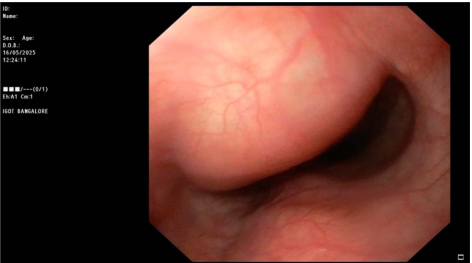
Esophagogastroduodenoscopy demonstrated a smooth, well-defined submucosal bulge in the anterior wall of the esophagus, approximately 4 cm in size, located 25–30 cm from the upper incisors with normally appearing overlying mucosa (Figure 1).
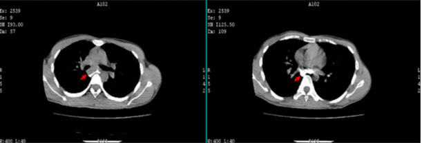
Contrast-enhanced computed tomography (CECT) of the chest revealed a smooth submucosal soft tissue lesion in the anterior wall of the esophagus measuring 2.5 cm × 4 cm at the level of D5 in the esophagus (Figure 2).
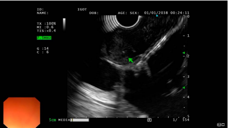
Endoscopic ultrasound (EUS) showed a homogeneous hypoechoic lesion arising from the muscularis propria with smooth margins and focal specks of calcification, measuring approximately 4 cm × 2.5 cm, extending from 25 to 29 cm from the incisors (Figure 3). The differential diagnosis included leiomyoma, gastrointestinal stromal tumor, and schwannoma. The absence of heterogeneity, cystic spaces, irregular margins, or adjacent lymphadenopathy favored a benign smooth muscle tumor, most consistent with leiomyoma. In view of these findings and the presence of significant dysphagia with associated weight loss, EUS-guided tissue acquisition was not pursued, and STER was planned for definitive management.
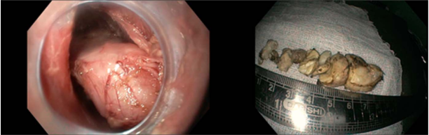
The procedure was performed using an Olympus 190 series gastroscope with a transparent cap (GIF-XP180N; Olympus, Tokyo, Japan). Prophylactic intravenous antibiotics (piperacillin-tazobactam) were administered before the procedure. After submucosal injection, a vertical mucosal incision was created from 19 to 21 cm from the incisors using Endocut Q mode (effect 2, cut duration 3, cut interval 3), and a submucosal tunnel was created. Submucosal dissection was performed using forced coagulation mode (effect 2, 40 W). A well-encapsulated submucosal lesion was identified, dissected from the underlying muscle layer, and resected piecemeal with a snare. Hemostasis was achieved using soft coagulation mode (effect 4, 80 W), and the mucosal defect was closed with hemoclips (Figure 4).
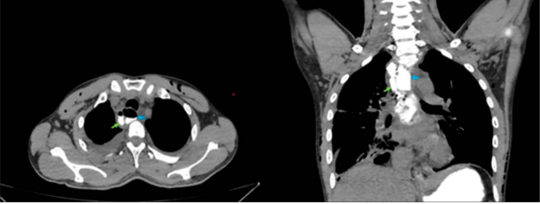
Twenty-four hours after procedure, the patient developed severe chest pain and fever spikes. On examination, he was tachycardic, with palpable subcutaneous crepitus suggestive of emphysema. Repeat CECT thorax revealed bilateral pneumothorax, subcutaneous emphysema, and circumferential wall thickening of the mid to lower esophagus, consistent with postprocedural changes. However, a small collection measuring 2.5 × 3 cm was noted adjacent to the STER site (Figure 5). No evidence of contrast leak was noted.
A repeat esophagogastroduodenoscopy demonstrated purulent discharge at the mucosal closure site between the hemoclips (Figure 6).
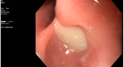
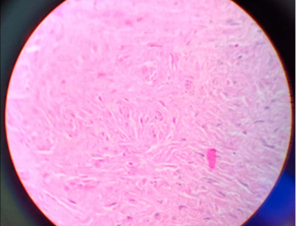
One hemoclip was removed to facilitate adequate drainage of pus. Empirical broad-spectrum antimicrobial therapy with meropenem was initiated for suspected deep mediastinal infection. A guided nasojejunal tube was placed for enteral nutrition, and analgesics were administered. Blood and pus cultures showed no microbial growth. Despite adequate endoscopic drainage and 5 days of meropenem, febrile episodes persisted, likely reflecting an ongoing inflammatory response and possible microresidual infection within the submucosal tunnel. After infectious disease consultation, antimicrobial therapy was escalated to teicoplanin and levofloxacin to enhance gram-positive and atypical coverage, after which the patient became afebrile within 4 days and demonstrating gradual clinical improvement. His oxygen requirement decreased, and he was successfully weaned off supplemental oxygen. Serial chest radiographs demonstrated resolution of pneumothorax and subcutaneous emphysema. Histopathological examination of the resected specimen revealed a well-circumscribed tumor composed of interlacing fascicles of bland smooth muscle cells. Mitotic figures were rare or absent, with no evidence of nuclear pleomorphism or necrosis. These morphological features were consistent with esophageal leiomyoma (Figure 7).
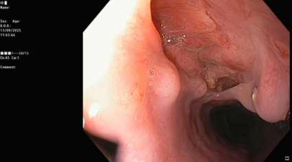
Follow-up contrast-enhanced CT thorax performed on postprocedure day 20, after clinical stabilization and resolution of fever, demonstrated complete resolution of the periesophageal collection. Subsequent upper gastrointestinal endoscopy revealed healthy granulation tissue with mucosal healing and pseudodiverticula at the prior STER site (Figure 8). After radiological and endoscopic confirmation of healing, the nasojejunal tube was removed, and the patient was discharged on oral antibiotics.
DISCUSSION
Esophageal leiomyomas are rare tumors, comprising less than 1% of gastrointestinal neoplasms. They are often asymptomatic but may present with dysphagia, weight loss, or chest discomfort when sufficiently large. Surgical resection, traditionally performed via thoracotomy or thoracoscopy, was the standard intervention but carried substantial morbidity. The development of minimally invasive endoscopic techniques—such as endoscopic submucosal dissection and STER—has transformed management by offering effective symptom relief with lower risk and faster recovery.
Although STER is generally safe, it is not without complications. Reported complications include bleeding, perforation, subcutaneous emphysema, and pneumothorax, most of which resolve with conservative management. However, the development of intramural or submucosal abscesses after STER is exceedingly rare. The mechanism typically involves bacterial contamination within the submucosal tunnel or incomplete closure of the mucosal entry site.
A report detailed a young woman who developed a submucosal tunnel abscess after STER for an esophageal schwannoma; endoscopy on postoperative day 7 revealed purulent collection, which was successfully managed by extending the mucosal incision and allowing natural drainage into the esophageal lumen, thereby avoiding surgery.4 Another case involved an 80-year-old woman who presented with dysphagia and fever due to a submucosal esophageal abscess that drained spontaneously; despite initial conservative antibiotic therapy, the condition progressed into intramural dissection, which was ultimately treated successfully with endoscopic septotomy.5 Similar experiences have also been reported after gastric endoscopic submucosal dissection, where a perigastric abscess developed secondary to delayed perforation. In that instance, the defect was intentionally left open to facilitate drainage into the gastric lumen, and the patient improved with antibiotics and conservative measures, avoiding surgical intervention.6
Management begins with a high index of suspicion and should be considered in patients who develop fever, chest discomfort, or retrosternal pain after STER. Treatment typically involves maintaining communication with the esophageal lumen to allow drainage, with debridement of pus and necrotic tissue within the tunnel if necessary. Broad-spectrum antibiotics, nutritional support via nasogastric or nasojejunal tube to bypass the affected segment, and early recognition remain the cornerstone of successful management.
Early esophageal wall abscess is a rare complication of STER. Prompt recognition and conservative management with antibiotics and controlled drainage can lead to full recovery and may obviate the need for surgery.
DISCLOSURES
Author contributions: All authors contributed to the conception and design of the study. Material preparation, data collection, and analysis were performed by PK Jain, RK Adi, and D. Saibaba. The first draft of the manuscript was written by AK Raghavendran. RP Desai, SA Vasu, and N. Patil contributed to data interpretation and critically reviewed the manuscript for important intellectual content. PK Jain provided overall supervision and approved the final version of the manuscript. All authors read and approved the final manuscript. PK Jain revised and finalized the manuscript and is the article guarantor.
Financial disclosure: None to report.
Informed consent was obtained for this case report.
Received November 24, 2025; Accepted January 5, 2026
REFERENCES
- Mathew G, Osueni A, Carter YM. Esophageal leiomyoma. In: StatPearls [Internet]. StatPearls Publishing, Treasure Island (FL), 2025. http://www.ncbi.nlm.nih.gov/books/NBK459298/
- Apdik T, Musharraf S, Gattuso P, Reddy V, Cheng L, Furci F. Leiomyomas of the gastrointestinal tract: A clinicopathologic review. Am J Clin Pathol. 2024;162(Supplement_1):S54.
- Isomoto H, Yamaguchi N, Minami H, Nakao K. Management of complications associated with endoscopic submucosal dissection/endoscopic mucosal resection for esophageal cancer. Dig Endosc. 2013;25(Supplement 1):29–38.
- Luo SB, Wang L, Liu ZQ, Li QL, Zhou PH. Endoscopic management of submucosal tunnel abscess following upper esophagus submucosal tunneling endoscopic resection. Endoscopy. 2025;57(S 01):E419–20.
- Jiao Y, Sikong YH, Zhang AJ, et al. Submucosal esophageal abscess evolving into intramural submucosal dissection: A case report. World J Clin Cases. 2022;10(29):10695–700.
- Nagae S, Kimoto Y, Sawada R, et al. Perigastric abscess caused by delayed perforation after gastric endoscopic submucosal dissection: Successful conservative treatment without perforation closure: A case report. J Med Case Rep. 2023;17(1):92.
ACG Case Rep J 2026;13:e01993. doi:10.14309/crj.0000000000001993. Published online: February 13, 2026.
Correspondence: Rakesh Kumar Adi, MBBS, MD, DM (rakeshkumaradi@yahoo.co.in).
Copyright: © 2026 The Author(s). Published by Wolters Kluwer Health, Inc. on behalf of The American College of Gastroenterology. This is an open access article distributed under the terms of the Creative Commons Attribution-Non Commercial-No Derivatives License 4.0 (CC BY-NC-ND), where it is permissible to download and share the work provided it is properly cited. The work cannot be changed in any way or used commercially without permission from the journal.